J Cerebrovasc Endovasc Neurosurg > Epub ahead of print
Youn, Park, and Kim: Coil embolization and recurrence of ruptured aneurysm originating from hyperplastic anterior choroidal artery

Abstract

Hyperplastic anterior choroidal artery (AchA) is an extremely rare congenital vascular variant that can be mistaken for other cerebral arteries. This case report presents a 38-year-old man who presented with a severe sudden-onset headache and was diagnosed with a ruptured aneurysm originating from a hyperplastic AchA. The aneurysm was successfully treated with coil embolization, but recurrence was detected after eight months, leading to additional surgical intervention. The discussion highlights the classification of hyperplastic AchA and emphasizes the importance of recognizing this anatomical variant to avoid complications during treatment. This case report underscores the need for awareness and understanding of hyperplastic AchA in the management of cerebral aneurysms.

INTRODUCTION

Hyperplastic anterior choroidal artery (AchA) is an extremely rare congenital vascular variant, first described in 1907 [2] and incidence reported between 1.8-2.3% [11,28]. Hyperplastic-AchA (H-AchA) is occasionally confused as ‘duplicated posterior cerebral artery (PCA)’ or ‘accessary PCA’ and associated with ‘fetal type PCA’ [26]. As H-AchA has both choroidal branches and communicating functions and it supplies a broader area that is usually served by the PCA compared to the normal AchA, this variant is often misdiagnosed as a duplicated or fetal type PCA.

CASE DESCRIPTION

A 38-year-old man presented with a severe sudden-onset headache and had no significant previous or familial medical history. Initial non-contrast computed tomography (CT) revealed subarachnoid hemorrhage (SAH) in the bilateral Sylvian fissure, peri-mesencephalic region, and basal cisterns (Fig. 1A). Subsequent CT angiography showed a suspicious aneurysm protruding superiorly from the left distal internal carotid artery (ICA) (Fig. 1B). Transfemoral cerebral angiography (TFCA) demonstrated that the aneurysm originated from the distal ICA. Initially, the origin of the aneurysm was considered to be a duplicated Posterior communicating artery (Pcom) or accessory PCA, but its proximal course followed the usual path of the typical cisternal segment of the AchA. Left vertebral angiograms (Alcock test) confirmed that the aneurysm originated more distally in the ICA, beyond the origin of the Pcom (Fig. 2A, B), and was definitively identified as an H-AchA aneurysm. The diameter of the H-AchA was even larger than that of the Pcom and had a lengthy course, extending distally to cover the posterior temporal area.
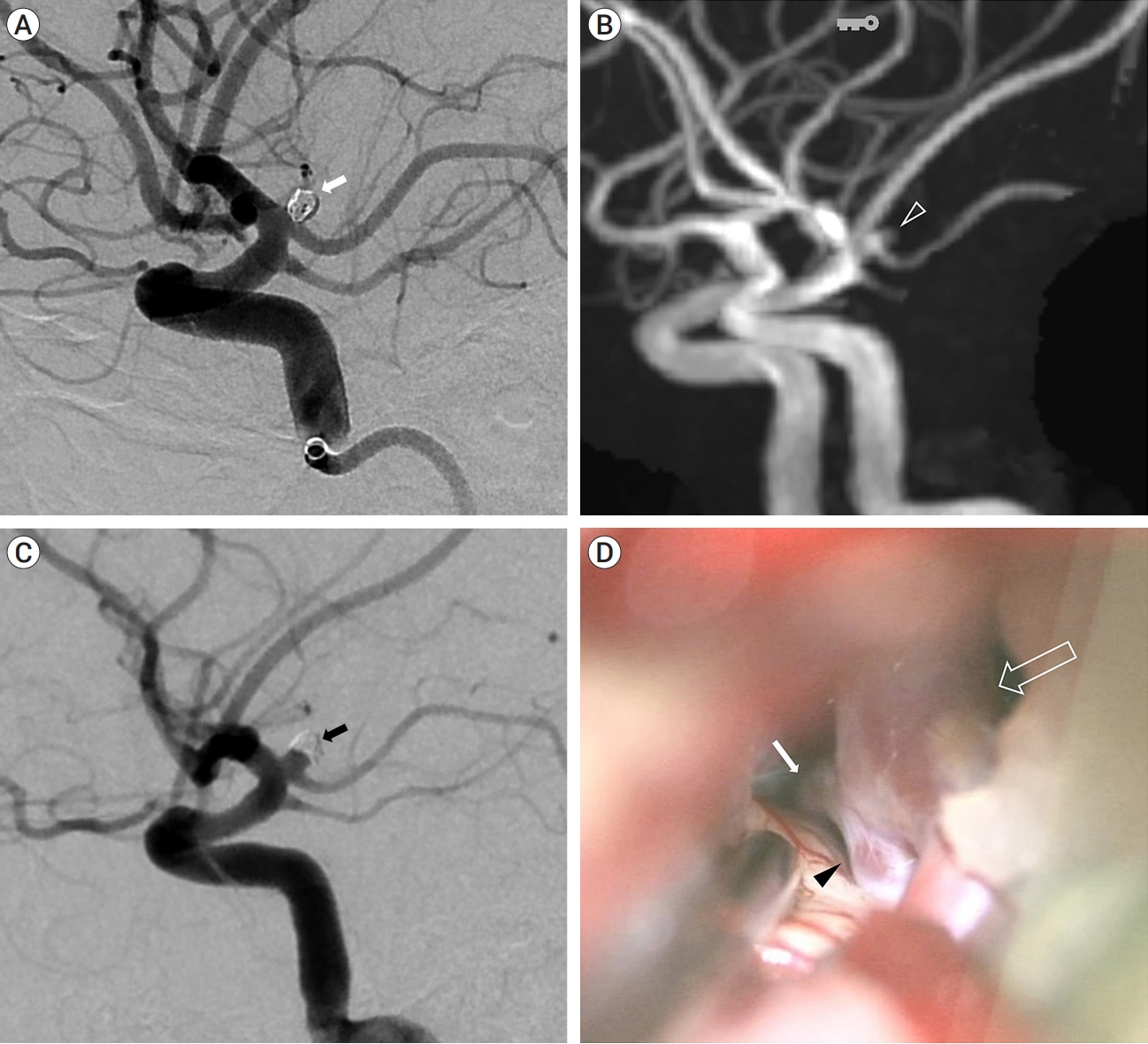
The aneurysm had a daughter sac and measured approximately 3.1 mm in diameter and it was treated with simple coil embolization. Under general anesthesia, through the right femoral artery, 6-Fr Shuttle and Sofia intermediate catheter were introduced into the left ICA. Then we used an Excelsior SL-10 microcatheter (Stryker Neurovascular, Kalamazoo, MI, USA) to navigate into the aneurysm sac and 2 detachable coils (Micrusframe S 3×5.4, Target nano 1.5×2, total coil length 7.4 cm) were filled into the sac. Immediate postprocedural angiograms showed near complete occlusion (Fig. 3A) and the packing density was about 24%. We tried additional coil (target nana 1×1) to improve packing density but failed. The patency of the fetal-type PCA and the hyperplastic AchA were preserved. The patient recovered well and was discharged after a few days without any significant events.
However, during regular follow-up observation, aneurysmal recurrence was detected on the magnetic resonance angiography (MRA) of pointwise encoding time reduction with radical acquisition (PETRA) sequence after eight months (Fig. 3B). TFCA confirmed the recurrence of the aneurysmal neck with coil compaction (Fig. 3C), and the patient ultimately underwent additional clipping surgery (Fig. 3D).

DISCUSSION

AchA aneurysms represent 2-4% of all intracranial aneurysms [24] and aneurysms associated with H-AchA have higher incidence [30] of 4-17% compared to typical AchA aneurysms. In a domestic study, out of 90 AchA aneurysms, 6 aneurysms (7%) were reported to be associated with H-AchA [13].
H-AchA was first classified into four types by Takahashi in 1990 based on the location of the remnant anastomosis between AchA and Pcom with PCA [30]. These types include hypertrophic uncal branch (type 1), anomalous temporal artery (type 2), anomalous occipito-parietal artery (type 3), and anomalous temporo-occipito-parietal artery (type 4). The patient in our case corresponds to type 2 of this classification, where H-AchA supplies some posterior temporal branches while the PCA proper gives rise to its own uncal branch. In our case, CT angiography revealed an additional artery arising from the communicating segment of the ICA, distal to the origin of the Pcom, which coursed postero-laterally. TFCA confirmed it as H-AchA and demonstrated its branches supplying the temporal areas along with the normal uncal branch of the PCA (Fig. 2).
H-AchA is referred to as ‘duplication of PCA’ or ‘true fetal PCA’, because it is believed to have similar developmental origins [9]. These terms highlight the resemblance and shared underlying causes of these vascular anomalies. Around the choroidal stage of fetal development (approximately at 5 weeks), the AchA initially supplies the territory of the PCA. As the brain volume increases and the posterior circulation develops, the normal pattern involves a reduction in blood supply from the AchA, leading to diminished hemispheric involvement of the AchA. However, in some cases, an unusual vascular regression occurs, resulting in a variant where the AchA continues to supply the posterior circulation from the anterior circulation and maintains its communicating function.
There are several cases about H-AchA aneurysm (Table 1). Present case is the first report of ruptured aneurysm from type 2 H-AchA and early recurrence of a ruptured H-AchA aneurysm after treatment. Previous studies have demonstrated that predisposing factors for recurrent coiled aneurysms include large side, widenecked, and ruptured [12,15]. Specially, large Pcom or fetaltype Pcom has been reported to be correlated with recurrence after coil embolization [5,14,16]. Considering that the location of the aneurysm in this case was almost identical to that of the Pcom, the aneurysm was ruptured, had a wide neck, and was associated with a large H-AchA, indicating a high risk of recurrence after coil embolization [3,4,16,23]. Further, the recurrence was somewhat earlier than usual aneurysms, and the immediate post-coil angiogram was close to Raymond class I [17,25], but the packing density was relatively low as 24%, which might have attributed to early recurrence [8,19,31]. When coiling a ruptured aneurysm in the ICA, considering early recurrence, a short term follow-up within 6 months would be recommended [6,22,23].

CONCLUSIONS

Although hyperplasia of AchA is a rare variant, it is associated with a relatively higher incidence of aneurysm formation compared to the original AchA. Given that the thromboembolic event (including sacrifice) of an H-AchA aneurysm can lead to more severe complications than in the case of the Pcom, it is important to recognize the anatomical variant of H-AchA. The recognition of this anatomical variant is crucial because the treatment-related complications of H-AchA aneurysms can result in severe and unexpected consequences [10]. Therefore, awareness of this variant is essential for providing appropriate and targeted care to patients.

NOTES

Disclosure

The authors report no conflict of interest concerning the materials or methods used in this study or the findings specified in this paper.

Fig. 1.
(A) Brain computed tomography (CT) reveals subarachnoid hemorrhage (SAH) within the bilateral sylvian fissure, perimesencephalic region, and basal cisterns. (B) CT angiography shows an aneurysm protruding superiorly from the left distal internal carotid artery (ICA) (open arrow).
jcen-2023-e2023-08-003f1.jpg
Fig. 2.
(A) Vertebral angiography with the Alcock test demonstrates a normal posterior communicating artery (Pcom) (black arrow). The aneurysm (open arrow) originates from the anomalous hyperplastic anterior choroidal artery (H-AchA) (white arrow), and some anomalous temporal branches are observed distally (open arrowhead). The posterior cerebral artery (PCA) follows its typical course, and the distal uncal branch is observed normally (white arrowhead). (B) The 3D rotation view shows the aneurysm (open arrow) originating from the H-AchA (white arrow), along with the distal normal uncal branch of the PCA (white arrowhead). Additionally, some anomalous temporal branches from the H-AchA are observed distally (open arrowhead).
jcen-2023-e2023-08-003f2.jpg
Fig. 3.
(A) The aneurysm was treated with simple coil embolization (white arrow). (B) After 8 months, magnetic resonance angiography (MRA) reveals the recurrence of the coiled aneurysm (open arrowhead). (C) Transfemoral cerebral angiography (TFCA) confirmed the recurrence of the aneurysm with coil compaction (black arrow). (D) Recurred aneurysm (black arrowhead) is seen between ICA (open arrow) and H-AchA (white arrow). Finally, surgical clipping was performed at the recurrent aneurysm. ICA, internal carotid artery; H-AchA, hyperplastic anterior choroidal artery
jcen-2023-e2023-08-003f3.jpg
Table 1.
Cases of H-AchA aneurysms reported to date
Year Authors Sex/Age Rupture/Unrupture Takahashi type Treatment
1990 Takahashi S et al. [30] n/a Ruptured Type 3 Clip
1999 Abrahams JM et al. [1] F/37 Unruptured Type 3 Clip
2000 Matsumoto K et al. [18] F/55 Unruptured Type 3 Clip
2005 Shioya H et al. [29] F/64 Ruptured Type 3 Clip
2011 Okazaki T et al. [21] M/18 Ruptured Type 3 Coil
2014 Hyun DK et al. [10] M/35 Ruptured Type 3 Coil
2018 Doi K et al. [7] n/a Unruptured Type 3 n/a
2019 Rutledge C et al. [27] F/45 Unruptured Type 4 Clip
2019 Mitsuhashi T et al. [20] M/48 Ruptured Type 3 Coil
2021 Present case M/39 Ruptured Type 2 Coil → Clip

AchA, hyperplastic anterior choroidal artery

REFERENCES

1. Abrahams JM, Hurst RW, Bagley LJ, Zager EL. Anterior choroidal artery supply to the posterior cerebral artery distribution: Embryological basis and clinical implications. Neurosurgery. 1999 Jun;44(6):1308-14.
crossref pmid
2. Blackburn IW. Anomalies of the encephalic arteries among the insane. A study of the arteries at the base of the encephallon in two hundred and twenty consecutive cases of mental disease, with special reference to anomalies of the circle of Willis. Journal of Comparative and Psychology. 1907 Nov;17(6):493-517.
crossref
3. Campi A, Ramzi N, Molyneux AJ, Summers PE, Kerr RS, Sneade M, et al. Retreatment of ruptured cerebral aneurysms in patients randomized by coiling or clipping in the International Subarachnoid Aneurysm Trial (ISAT). Stroke. 2007 May;38(5):1538-44.
crossref pmid
4. Choi DS, Kim MC, Lee SK, Willinsky RA, Terbrugge KG. Clinical and angiographic long-term follow-up of completely coiled intracranial aneurysms using endovascular technique. J Neurosurg. 2010 Mar;112(3):575-81.
crossref pmid
5. Choi HH, Cho YD, Yoo DH, Lee HS, Kim SH, Jang D, et al. Impact of fetal-type posterior cerebral artery on recanalization of posterior communicating artery aneurysms after coil embolization: Matched-pair case-control study. J Neurointerv Surg. 2020 Aug;12(8):783-7.
crossref pmid
6. Cognard C, Weill A, Spelle L, Piotin M, Castaings L, Rey A, et al. Long-term angiographic follow-up of 169 intracranial berry aneurysms occluded with detachable coils. Radiology. 1999 Aug;212(2):348-56.
crossref pmid
7. Doi K, Mizuno T, Shigematsu Y, Kobayashi O, Takezaki T, Muta D, et al. A new type of hyperplastic anterior choroidal artery. J Clin Neurosci. 2018 May;51:72-4.
crossref pmid
8. Gaba RC, Ansari SA, Roy SS, Marden FA, Viana MA, Malisch TW. Embolization of intracranial aneurysms with hydrogel-coated coils versus inert platinum coils: Effects on packing density, coil length and quantity, procedure performance, cost, length of hospital stay, and durability of therapy. Stroke. 2006 Jun;37(6):1443-50.
crossref pmid
9. Hahm MH, Moon S. Anomalous hyperplastic anterior choroidal artery and its mimicker: A case series. Surg Radiol Anat. 2021 Dec;43(12):1961-5.
crossref pmid pdf
10. Hyun DK, Shim YS, Park HS, Oh SY. Thromboembolic complication following neurointervention in ruptured anomalous hyperplastic anterior choroidal artery aneurysm. Neuroradiol J. 2014 Feb;27(1):103-7.
crossref pmid pmc pdf
11. Iihoshi S, Nonaka T, Miyata K, Houkin K. Angiographic analysis of variations and anomalous origin of the anterior choroidal artery and posterior communicating artery. No Shinkei Geka. 2010 38(6):523-30.
pmid
12. Jeon JP, Cho YD, Yoo DH, Moon J, Lee J, Cho WS, et al. Risk factor analysis of recanalization timing in coiled aneurysms: Early versus late recanalization. AJNR Am J Neuroradiol. 2017 Sep;38(9):1765-70.
crossref pmid pmc
13. Kang HS, Kwon BJ, Kwon OK, Jung C, Kim JE, Oh CW, et al. Endovascular coil embolization of anterior choroidal artery aneurysms. Clinical article. J Neurosurg. 2009 Nov;111(5):963-9.
pmid
14. Kim MJ, Chung J, Park KY, Kim DJ, Kim BM, Suh SH, et al. Recurrence and risk factors of posterior communicating artery aneurysms after endovascular treatment. Acta Neurochir (Wien). 2021 Aug;163(8):2319-26.
crossref pmid pdf
15. Koyanagi M, Ishii A, Imamura H, Satow T, Yoshida K, Hasegawa H, et al. Long-term outcomes of coil embolization of unruptured intracranial aneurysms. J Neurosurg. 2018 Dec;129(6):1492-8.
crossref pmid
16. Lee HJ, Choi JH, Shin YS, Lee KS, Kim BS. Risk factors for the recurrence of posterior communicating artery aneurysm: The significance of fetal-type posterior cerebral artery. J Stroke Cerebrovasc Dis. 2021 Jul;30(7):105821.
crossref pmid
17. Mascitelli JR, Moyle H, Oermann EK, Polykarpou MF, Patel AA, Doshi AH, et al. An update to the Raymond-Roy Occlusion Classification of intracranial aneurysms treated with coil embolization. J Neurointerv Surg. 2015 Jul;7(7):496-502.
crossref pmid
18. Matsumoto K, Akagi K, Abekura M, Ohkawa M, Tasaki O, Oshino S. Cerebral aneurysm associated with an anomalous hyperplastic anterior choroidal artery. Acta Neurochir (Wien). 2000 142(3):347-50.
crossref pmid pdf
19. Mehra M, Hurley MC, Gounis MJ, King RM, Shaibani A, Dabus G, et al. The impact of coil shape design on angiographic occlusion, packing density and coil mass uniformity in aneurysm embolization: An in vitro study. J Neurointerv Surg. 2011 Jun;3(2):131-6.
crossref pmid
20. Mitsuhashi T, Oishi H, Teranishi K, Kodama T, Kudo K, Tokugawa J, et al. Ruptured anomalous hyperplastic anterior choroidal artery aneurysm: A case report. Br J Neurosurg. 2019 Jun;37(3):296-7.
crossref pmid
21. Okazaki T, Shibukawa M, Kiura Y, Sakamoto S, Ichinose N, Eguchi K, et al. Endovascular coil embolization for ruptured aneurysm associated with persistent primitive anterior choroidal artery--Case report. Neurol Med Chir (Tokyo). 2011 51(2):127-9.
crossref pmid
22. Plowman RS, Clarke A, Clarke M, Byrne JV. Sixteen-year single-surgeon experience with coil embolization for ruptured intracranial aneurysms: Recurrence rates and incidence of late rebleeding. Clinical article. J Neurosurg. 2011 Mar;114(3):863-74.
pmid
23. Raymond J, Guilbert F, Weill A, Georganos SA, Juravsky L, Lambert A, et al. Long-term angiographic recurrences after selective endovascular treatment of aneurysms with detachable coils. Stroke. 2003 Jun;34(6):1398-403.
crossref pmid
24. Fujii K, Fradd B. Microsurgical anatomy of the anterior choroidal artery. Surg Neurol. 1979 Aug;12(2):171-87.
pmid
25. Roy D, Milot G, Raymond J. Endovascular treatment of unruptured aneurysms. Stroke. 2001 Sep;32(9):1998-2004.
crossref pmid
26. Rusu MC, Vrapciu AD, Lazar M. A rare variant of accessory posterior cerebral artery. Surg Radiol Anat. 2023 May;45(5):523-6.
crossref pmid pdf
27. Rutledge C, Jonzzon S, Andrew Winkler E, Hetts SW, Abla AA. Transcortical transventricular transchoroidal-fissure approach to distal fusiform hyperplastic anterior choroidal artery aneurysms. Br J Neurosurg. 2019 Aug;37(4):723-7.
crossref pmid
28. Shah AU, Joshi AR, Rai PR, Kapse P. Hyperplastic anterior choroidal artery-A rare variant detected on MR angiography. Indian J Radiol Imaging. 2022 Jun;32(2):266-9.
crossref pmid pmc
29. Shioya H, Kikuchi K, Suda Y, Shoji H, Shindo K. Ruptured internal carotid -posterior communicating artery aneurysm associated with an anomalous hyperplastic anterior choroidal artery and aneurysm: Case report. No Shinkei Geka. 2005 Feb;33(2):155-62.
pmid
30. Takahashi S, Suga T, Kawata Y, Sakamoto K. Anterior choroidal artery: Angiographic analysis of variations and anomalies. AJNR Am J Neuroradiol. 1990 Jul-Aug;11(4):719-29.
pmid pmc
31. Tian Z, Liu J, Zhang Y, Zhang Y, Zhang X, Zhang H, et al. Risk factors of angiographic recurrence after endovascular coil embolization of intracranial saccular aneurysms: A retrospective study using a multicenter database. Front Neurol. 2020 Sep;11:1026.
crossref pmid pmc
TOOLS
METRICS Graph View
  • 0 Crossref
  •  0 Scopus
  • 786 View
  • 20 Download
ORCID iDs

Sang Kyu Park
https://orcid.org/0000-0001-9231-0716

Related articles


ABOUT
BROWSE ARTICLES
EDITORIAL POLICY
FOR CONTRIBUTORS
Editorial Office
The Journal of Cerebrovascular and Endovascular Neurosurgery (JCEN), Department of Neurosurgery, Wonkwang University
School of Medicine and Hospital, 895, Muwang-ro, Iksan-si, Jeollabuk-do 54538, Korea
Tel: +82-2-2279-9560    Fax: +82-2-2279-9561    E-mail: editor.jcen@the-jcen.org                

Copyright © 2024 by Korean Society of Cerebrovascular Surgeons and Korean NeuroEndovascular Society.

Developed in M2PI

Close layer
prev next