J Cerebrovasc Endovasc Neurosurg > Volume 26(1); 2024 > Article
Eraky, Feller, Tolba, Porwal, Sacho, and Hedayat: Flow diverter stenting for intracranial aneurysms in the pediatric population: Two case reports and literature review

Abstract

The Pipeline Embolization Device (PED) is a flow-diverting intraluminal device that is approved for use in adults 18 years or older with internal carotid artery aneurysms. However, it can also be used off-label in pediatric patients with aneurysms that cannot be resolved with traditional endovascular treatments. Herein, we present two cases of flow diversion in the pediatric population with complete obliteration of the aneurysm and excellent outcomes. Flow diversion has been shown to be a safe endovascular option in treating complex aneurysms in children. Larger-sized, multicenter trials are encouraged to compare outcomes between flow diversion and other aneurysm treatment options given the rarity of pediatric aneurysms.

INTRODUCTION

Intracranial aneurysms (IAs) are characteristically rare in the pediatric population and their treatment is often more difficult and complex than that of adults [18]. Additionally, in the pediatric population, ruptured aneurysms are more common than unruptured ones [20]. Management options for pediatric IAs range from observation with serial follow-up imaging to microsurgical or endovascular intervention [15].
Endovascular coil embolization involves placing coils, densely packed inside the aneurysm to induce thrombosis and occlusion. The Pipeline Embolization Device (PED) is a flow-diverting intraluminal device that induces thrombosis and occlusion of the aneurysm with reendothelialization of the device [15,27,30]. The device is approved for use in adults 18 years or older with internal carotid artery (ICA) aneurysms arising from the petrous segment to the terminus. However, it can also be used off-label in pediatric patients with aneurysms that cannot be resolved with traditional endovascular treatments [26]. During flow diversion, a microcatheter is navigated past the aneurysm in the native vessel (without needing to enter the aneurysm). The PED is then deployed across the neck of the aneurysm in the parent blood vessel. If successful, the aneurysm should completely thrombose and occlude in the following 6 weeks to 6 months [6,26].

CASE DESCRIPTION

First case

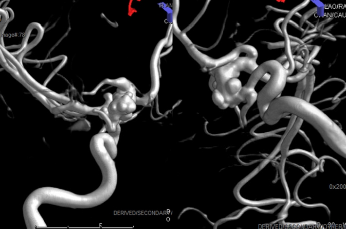
A 15-year-old girl with a sibling affected by a vascular anomaly presented to neurosurgery clinic following incidental discovery of a left-sided vascular abnormality during imaging after sustaining a concussion. Since her concussion three weeks prior, she had been complaining of post-concussive symptoms as well as intermittent bilateral visual blurriness. Magnetic resonance angiography (MRA) showed a dysplastic, fusiform aneurysmal dilatation of the left supraclinoid terminal carotid artery with some compression of the adjacent chiasm and left optic nerve. The aneurysm measured 11 mm×9 mm via catheter angiography (Fig. 1). Additionally, multiple secondary daughter sacs arose from the supraclinoid and communicating segment of the ICA to the carotid terminus with aneurysmal incorporation of the proximal M1 segment. The anterior choroidal artery with fusiform, dysplastic morphology originated from the dome of the aneurysm.
It was determined that this aneurysm was concerning for risk of rupture given the dysplastic, fusiform shape. Flow diversion stent placement via right common femoral artery approach was offered off-label. Under fluoroscopic magnification and roadmap imaging, a microcatheter was advanced through the ICA and into the frontal M2 branch, and wire was removed. Under continuous fluoroscopic guidance, a 4.75 by 35 mm Pipeline Flow Diverting stent was then deployed from the ophthalmic segment of the ICA to the middle of the M1 segment of the left middle cerebral artery, ensuring that the device was completely expanded and well opposed. Fluoroscopic images determined good wall apposition and final angiographic images demonstrated stagnant flow within the aneurysmal daughter sacs with preserved, albeit slower, flow within the anterior cerebral artery as well as the ophthalmic artery. There was well-maintained flow within the anterior choroidal artery.
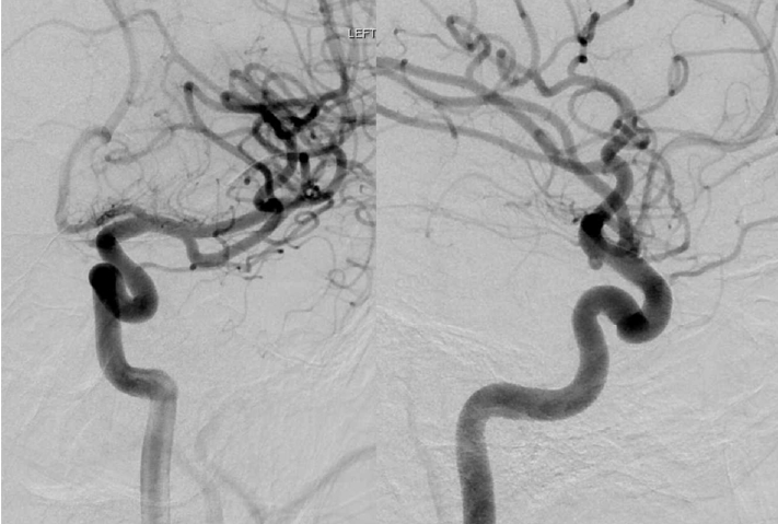
Two months post-flow diverter stenting, the patient had resolution of headaches and no new neurological symptoms. MRA showed continued diminishment of aneurysm filling, encouraging for aneurysm exclusion. A 6-month follow-up catheter angiogram showed near complete resolution of the left terminal ICA aneurysm; however, evidence of non-flow-limiting tapering stenosis of the left supraclinoid carotid was noted and has continued to be monitored (Fig. 2).

Second case

A 15-year-old boy with past medical history of Juvenile myoclonic epilepsy (JME) as a sequela of viral meningitis two years prior was admitted to the hospital for evaluation after changes in seizure semiology. During this admission, his MRI (magnetic resonance imaging) brain showed a suspected left supra-clinoid carotid artery aneurysm that was further confirmed using MRA. Subsequently, a catheter angiogram revealed a dysplastic segment of the left-sided supraclinoid ICA with a broad-based, saccular 6.2 mm aneurysm and a 2 mm aneurysm arising on the opposite wall of the ICA (Fig. 3).
After discussing risks and benefits, family agreed with PED as a treatment for the left ICA aneurysm. Under a traditional fluoroscopic roadmap, a PED of 4.72 mm×14 mm was subsequently deployed uneventfully with good wall apposition and coverage of the aneurysm extending from just beyond the ophthalmic artery proximally to just proximal to the origin of the posterior communicating artery distally.
Three months after the procedure, the patient obtained a repeat MRA that showed partial obliteration of the aneurysm with a thrombus noted in the aneurysm dome. A six-month follow-up catheter angiogram showed complete thrombosis of the aneurysm and patent PED in the left ICA (Fig. 4). The patient continued to follow up in the clinic and at 18 months after PED placement, MRA showed no evidence of aneurysmal recurrence; he continues to do well clinically.

DISCUSSION

Aneurysm characteristics in the pediatric population, such as large size, fusiform shape, or dissecting vessel walls make them difficult to treat using conventional microsurgical treatment or endovascular options. This is in part due to the fact that both the blood vessels and the surgical field are smaller in the pediatric population. The pediatric population is also more sensitive to blood loss [6,15],18-20],25]. Thus, choosing the appropriate treatment option between endovascular and microsurgery is controversial; there remains no protocol for IA management in the pediatric population [20]. Despite this, with treatment for aneurysms shifting from microsurgery to endovascular methods in adults, this trend has started to extend to the pediatric population as well [10,35].
Some endovascular options, such as coiling have shown significant rates of recanalization and high mortality and morbidity rates in the general population [16]. Recently, flow diverters have shifted from on-label use in ICA aneurysms only to distal and posterior circulation aneurysms as well [11,16,17,26,31,36,37]. Moreover, flow diverter use has expanded to treat ruptured aneurysms in the acute phase with favorable outcomes despite the need for dual antiplatelet therapies [12]. Flow diverters work by inducing vessel reconstruction. As the parent vessel’s endothelial layer starts remodeling, the aneurysm becomes isolated from the circulation; thus, creating aneurysmal flow disruption and aneurysm thrombosis [8]. The use of flow diverters in the pediatric population is limited to a small number of cases reported in the literature (Table 1) and discussed further below with each underlying etiological aneurysm subtype.
While children do not have chronic exposure to the environmental factors or medical comorbidities associated with aneurysm development as with adults, up to 30% of the pediatric population has underlying conditions related to aneurysm development and growth. These include polycystic kidney disease, Ehlers-Danlos syndrome, fibromuscular dysplasia, infectious endocarditis, and trauma [27,30,32]. Both of our patients did not undergo genetic evaluation, however, given that the first patient has a sibling with a vascular anomaly, he may have had a genetic predisposition.
One etiology of pediatric aneurysms is trauma. Traumatic aneurysms occur due to penetrating or closed trauma of the head [34]. In contrast to adults, the incidence of traumatic aneurysms in pediatrics is very high, reaching 39% in some studies [20]. Furthermore, aneurysm recurrence risk, aneurysmal growth rate, and rupture incidence are higher in the pediatric population, thus raising the importance of early treatment over just observation [20,33]. Similarly, dissecting aneurysms are more common in children, and in contrast to arterial dissection that can heal alone or with medications, dissecting aneurysms tend to grow, thus warranting treatment [13].
Infectious aneurysms, on the other hand, result from the inflammatory reaction to an infectious agent within the adventitia and then muscularis layer, resulting in a very friable, thin-walled aneurysm. Typically these aneurysms appear without a distinguishable neck, so they are considered pseudoaneurysms [14,28]. About 15% of IAs in pediatrics are infectious [21]. Due to their histopathology, infectious aneurysms in pediatric patients have a high risk of rupture along with a latency of complete obliteration. It is additionally challenging to treat infectious aneurysms using flow diversion given the nature of placing an endovascular device in the context of potential bacteremia [4]. Yet, many studies have shown success using flow diverters to treat infectious aneurysms in the pediatric population. Ares et al. reported using PED to treat a two-year-old male with an infectious basilar apex aneurysm; 6-month angiography showed complete obliteration [4]. Another case reported by Appelboom et al. showed complete occlusion of a ten-year-old female’s infectious cavernous carotid aneurysm treated with a flow diverter [3]. A third case reported by Samples et al. showed a 10-year-old female with two reputed, infectious, MCA (middle cerebral artery) aneurysms. The proximal one was treated with coil embolization and the distal one was treated with a flow diverter. Again, 6-month angiogram showed complete obliteration of both aneurysms [29].
While seen to be successful in many case reports, there are four main potential problems related to using flow diverters in pediatrics. First, antiplatelet treatment prior to flow diverters is essential to decrease peri- and post-procedural thromboembolic events [2]. However, due to ADP-induced hyporeactivity, Aspirin-induced Reye syndrome, possible hyper response to antiplatelets and the absence of standard guidelines, using antiplatelets and adjusting their doses is challenging in pediatric patients; additionally, antiplatelet administration is extremely variable [5,7,9]. In a trial studying the appropriate antiplatelet doses in children having heart disease, Li et al. found that to achieve a sufficient therapeutic effect, 0.2 mg/kg of clopidogrel may be enough [23]. The second potential problem of utilizing flow diverters in the pediatric population is that the continuous growth of children raises concerns regarding the presence of a foreign body inside the growing blood vessels and potential long-term complications [5]. Third, the lack of experience in using flow diverters in the pediatric population can pose technical challenges for the operator [22]. Fourth, flow diverters can theoretically increase the risk of developing perforator strokes, particularly after dual antiplatelets are de-escalated to a single antiplatelet [1,24].

CONCLUSIONS

Flow diversion has been shown to be a safe endovascular option in treating complex aneurysms in children and can lead to complete occlusion with excellent outcomes. Larger-sized, multicenter trials are encouraged to compare outcomes between flow diversion and other aneurysm treatment options given the rarity of pediatric aneurysms.

NOTES

Disclosure

The authors report no conflict of interest concerning the materials or methods used in this study or the findings specified in this paper.

Fig. 1.
A 3D reconstruction angiogram reveals a dysplastic, fusiform, multilobular aneurysm measuring 11 mm×9 mm. 3D, threedimensional
jcen-2023-e2023-04-001f1.jpg
Fig. 2.
Six-month follow-up angiogram shows complete resolution of the left terminal ICA aneurysm and non-flow-limiting tapering stenosis of the left supraclinoid carotid. ICA, internal carotid artery
jcen-2023-e2023-04-001f2.jpg
Fig. 3.
A 3D reconstruction angiogram reveals a dysplastic segment of the left-sided supraclinoid ICA with a broad-based, saccular 6.2 mm aneurysm. 3D, three-dimensional; ICA, internal carotid artery
jcen-2023-e2023-04-001f3.jpg
Fig. 4.
Six-month follow-up angiogram shows complete thrombosis of the left ICA aneurysm. ICA, internal carotid artery
jcen-2023-e2023-04-001f4.jpg
Table 1.
Summary of pediatric intracranial aneurysms treated with flow diversion
Author and year Ref. N. of PEDs Aneurysm locations Age (years) Sex Aneurysm features
Kumaria et al. 2021 [22] 1 ICA 8 1 male Serpiginous
Colby et al. 2018 [10] 1 MCA 0.75 1 female Saccular
Mohammad et al. 2017 [26] 1 Basilar 15 1 male Partially thrombosed aneurysm
Vachhani et al. 2016 [35] 1 A2 12 1 female Fusiform
Vargas et al. 2016 [36] 1 A2 8 1 male Saccular
2 Basilar, ICA 9 1 male Fusiform
Kan et al. 2016 [19] 1 Basilar 16 1 male Fusiform
Takemoto et al. 2014 [32] 2 ICA, Acom N/A N/A N/A
Burrows et al. 2013 [6] 1 M2 15 1 male Fusiform
Saraf et al. 2012 [30] 1 Basilar N/A N/A Partially thrombosed aneurysm
Crowley et al. 2009 [11] 1 Basilar 1.8 1 male Fusiform
Ares et al. 2019 [4] 1 Basilar 2 1 male Infectious, Sub occlusive thrombosed aneurysm
Tonetti et al. 2021 [33] 1 A2 15 1 male Fusiform, Partially thrombosed
Cherian et al. 2021 [7] 46 N/A Range (3-21) 27 male N/A
12 female
Samples et al. 2021 [29] 1 M1 10 1 female Ruptured, Infectious
Cunegatto-Braga 2018 [12] 1 PCA 4 1 male Ruptured, Dissecting
Ghali et al. 2018 [15] 1 ICA 13 1 male N/A
1 ACA 14 1 male Traumatic
1 Basilar 1.16 1 male Dissecting
Sastry et al. 2018 [31] 1 Basilar 13 1 male Iatrogenic
Navarro et al. 2015 [27] 1 Pcom 4 Male Saccular
2 Vertebrobasilar, ICA 11 1 female Saccular
Jia et al. 2020 [17] 1 Vertebrobasilar N/A 1 female Dissecting
Barburoglu and Arat 2017 [5] 7 (4) ICA, (1) A2, (1) M1, (1) V4 Range (3-16) 6 males N/A
1 female
Ikeda et al. 2015 [16] 1 M2 12 1 female Fusiform
Appelboom et al. 2010 [3] 1 ICA 10 1 female Infectious
Lin et al. 2016 [24] 1 A2 13 1 male Dissecting
3 M2 16 1 male Fusiform
De Barros et al. 2011 [13] 2 Basilar 12 1 female Dissecting
2 Basilar 5 1 female Dissecting
4 Vertebrobasilar 7 1 male Dissecting
Cinar et al. 2013 [8] 1 ICA 8 1 female Saccular
Zarzecka et al. 2014 [37] 2 V4 15 1 male Fusiform
Abla et al. 2014 [1] 1 ICA 10 1 male Fusiform
Trivelato et al. 2017 [34] 1 ICA 9 1 female Traumatic
Cobb et al. 2017 [9] 1 ICA 13 1 female Iatrogenic
Lubicz et al. 2010 [25] 1 Basilar 17 1 female Fusiform
1 ICA 10 1 female Saccular

Ref., reference number; N. of PEDs, number of pediatric patients; ICA, internal cerebral artery; MCA, middle cerebral artery; A2, second segment of the anterior cerebral artery; Acom, anterior communicating artery; M1, first segment of the middle cerebral artery; M2, second segment of the middle cerebral artery; PCA, posterior cerebral artery; ACA, anterior cerebral artery; Pcom, posterior communicating artery; V4, fourth segment of the vertebral artery

REFERENCES

1. Abla AA, Zaidi HA, Crowley RW, Britz GW, McDougall CG, Albuquerque FC, et al. Optic chiasm compression from mass effect and thrombus formation following unsuccessful treatment of a giant supraclinoid ICA aneurysm with the Pipeline device: Open surgical bailout with STA-MCA bypass and parent vessel occlusion. J Neurosurg Pediatr. 2014 Jul;14(1):31-7.
crossref pmid
2. Adeeb N, Griessenauer CJ, Foreman PM, Moore JM, Shallwani H, Motiei-Langroudi R, et al. Use of platelet function testing before pipeline embolization device placement: A multicenter cohort study. Stroke. 2017 May;48(5):1322-30.
crossref pmid
3. Appelboom G, Kadri K, Hassan F, Leclerc X. Infectious aneurysm of the cavernous carotid artery in a child treated with a new-generation of flow-diverting stent graft: Case report. Neurosurgery. 2010 Mar;66(3):e623-4; discussion e624.
pmid
4. Ares WJ, Tonetti DA, Greene S, Sharma MS, Xavier F, Jankowitz BT, et al. Pipeline embolization of an infectious basilar artery aneurysm in a 2-year-old child: Case report, discussion of the literature and perioperative considerations. Oper Neurosurg (Hagerstown). 2019 Nov;17(5):e224-8.
crossref pmid pdf
5. Barburoglu M, Arat A. Flow diverters in the treatment of pediatric cerebrovascular diseases. AJNR Am J Neuroradiol. 2017 Jan;38(1):113-8.
crossref pmid pmc
6. Burrows AM, Zipfel G, Lanzino G. Treatment of a pediatric recurrent fusiform middle cerebral artery (MCA) aneurysm with a flow diverter. J Neurointerv Surg. 2013 Nov;5(6):e47.
crossref pmid
7. Cherian J, Srinivasan V, Froehler MT, Grossberg JA, Cawley CM, Hanel RA, et al. Flow diversion for treatment of intracranial aneurysms in pediatric patients: Multicenter case series. Neurosurgery. 2020 Jul;87(1):53-62.
crossref pmid pdf
8. Cinar C, Bozkaya H, Oran I. Endovascular treatment of cranial aneurysms with the pipeline flow diverting stent: Preliminary midterm results. Diagn Interv Radiol. 2013 MarApr;19(2):154-64.
crossref pmid
9. Cobb MIH, Zomorodi AR, Hauck EF, Smith TP, Fernando Gonzalez L. Optimal pediatric dosing of anti-platelet agents for pipeline stent embolization: A case report and review of the literature. Childs Nerv Syst. 2017 Apr;33(4):685-90.
crossref pmid pdf
10. Colby GP, Jiang B, Bender MT, Beaty NB, Westbroek EM, Xu R, et al. Pipeline-assisted coil embolization of a large middle cerebral artery pseudoaneurysm in a 9-month-old infant: Experience from the youngest flow diversion case. J Neurosurg Pediatr. 2018 Nov;22(5):532-40.
crossref pmid
11. Crowley RW, Evans AJ, Kassell NF, Jensen ME, Dumont AS. Endovascular treatment of a fusiform basilar artery aneurysm using multiple “in-stent stents”: Technical note. J Neurosurg Pediatr. 2009 Jun;3(6):496-500.
pmid
12. Cunegatto-Braga M, Hogan B, Aguilar-Salinas P, Beier AD, Hanel RA. Pipeline embolization device flow diversion for a dissecting ruptured posterior cerebral artery aneurysm in a pediatric patient. World Neurosurg. 2018 Sep;117:255-60.
crossref pmid
13. de Barros Faria M, Castro RN, Lundquist J, Scrivano E, Ceratto R, Ferrario A, et al. The role of the pipeline embolization device for the treatment of dissecting intracranial aneurysms. AJNR Am J Neuroradiol. 2011 Dec;32(11):2192-5.
crossref pmid pmc
14. Eddleman CS, Surdell D, DiPatri A, Tomita T, Shaibani A. Infectious intracranial aneurysms in the pediatric population: Endovascular treatment with Onyx. Childs Nerv Syst. 2008 Aug;24(8):909-15.
crossref pmid pdf
15. Ghali MGZ, Srinivasan VM, Cherian J, Wagner KM, Chen SR, Johnson J, et al. Multimodal treatment of intracranial aneurysms in children: Clinical case series and review of the literature. World Neurosurg. 2018 Mar;111:e294-307.
crossref pmid
16. Ikeda DS, Marlin ES, Shaw A, Powers CJ. Successful endovascular reconstruction of a recurrent giant middle cerebral artery aneurysm with multiple telescoping flow diverters in a pediatric patient. Pediatr Neurosurg. 2015 50(2):88-93.
crossref pmid pdf
17. Jia L, Wang J, Zhang L, Zhang Y, You W, Yang X, et al. Pediatric patient with a giant vertebrobasilar dissecting aneurysm successfully treated with three pipeline embolization devices. Front Neurol. 2020 Jul;11:633.
crossref pmid pmc
18. Kalani MY, Elhadi AM, Ramey W, Nakaji P, Albuquerque FC, McDougall CG, et al. Revascularization and pediatric aneurysm surgery. J Neurosurg Pediatr. 2014 Jun;13(6):641-6.
crossref pmid
19. Kan P, Mokin M, Puri AS, Wakhloo AK. Successful treatment of a giant pediatric fusiform basilar trunk aneurysm with surpass flow diverter. J Neurointerv Surg. 2016 Jun;8(6):e23.
crossref pmid pmc
20. Kim M, Lee HS, Lee S, Park JC, Ahn JS, Kwon DH, et al. Pediatric intracranial aneurysms: Favorable outcomes despite rareness and complexity. World Neurosurg. 2019 May;125:e1203-16.
crossref pmid
21. Krings T, Geibprasert S, terBrugge KG. Pathomechanisms and treatment of pediatric aneurysms. Childs Nerv Syst. 2010 Oct;26(10):1309-18.
crossref pmid pdf
22. Kumaria A, McConachie NS, Macarthur DC. Successful treatment of giant cavernous carotid artery aneurysm in a child using a flow diverter stent. Br J Neurosurg. 2021 Feb;35(1):122-4.
crossref pmid
23. Li JS, Yow E, Berezny KY, Bokesch PM, Takahashi M, Graham TP, et al. Dosing of clopidogrel for platelet inhibition in infants and young children: Primary results of the Platelet Inhibition in Children On cLOpidogrel (PICOLO) trial. Circulation. 2008 Jan;117(4):553-9.
crossref pmid
24. Lin N, Lanzino G, Lopes DK, Arthur AS, Ogilvy CS, Ecker RD, et al. Treatment of distal anterior circulation aneurysms with the pipeline embolization device: A US multicenter experience. Neurosurgery. 2016 Jul;79(1):14-22.
pmid
25. Lubicz B, Collignon L, Raphaeli G, Pruvo JP, Bruneau M, De Witte O, et al. Flow-diverter stent for the endovascular treatment of intracranial aneurysms: A prospective study in 29 patients with 34 aneurysms. Stroke. 2010 Oct;41(10):2247-53.
crossref pmid
26. Mohammad LM, Coon AL, Carlson AP. Resolution of giant basilar artery aneurysm compression and reversal of sensorineural hearing loss with use of a flow diverter: Case report. J Neurosurg Pediatr. 2017 Jul;20(1):81-5.
crossref pmid
27. Navarro R, Brown BL, Beier A, Ranalli N, Aldana P, Hanel RA. Flow diversion for complex intracranial aneurysms in young children. J Neurosurg Pediatr. 2015 Mar;15(3):276-81.
crossref pmid
28. Peters PJ, Harrison T, Lennox JL. A dangerous dilemma: Management of infectious intracranial aneurysms complicating endocarditis. Lancet Infect Dis. 2006 Nov;6(11):742-8.
crossref pmid
29. Samples DC, Ravindra VM, Thoms DJ, Tarasiewicz I, Grandhi R. Successful flow diversion treatment of ruptured infectious middle cerebral artery aneurysms with the use of Pipeline Flex with Shield technology. Interv Neuroradiol. 2021 Apr;27(2):225-9.
crossref pmid pmc pdf
30. Saraf R, Shrivastava M, Siddhartha W, Limaye U. Intracranial pediatric aneurysms: Endovascular treatment and its outcome. J Neurosurg Pediatr. 2012 Sep;10(3):230-40.
crossref pmid
31. Sastry RA, Koch MJ, Grannan BL, Stapleton CJ, Butler WE, Patel AB. Flow diversion of a recurrent, iatrogenic basilar tip aneurysm in a pediatric patient: Case report. J Neurosurg Pediatr. 2018 Jan;21(1):90-3.
crossref pmid
32. Takemoto K, Tateshima S, Golshan A, Gonzalez N, Jahan R, Duckwiler G, et al. Endovascular treatment of pediatric intracranial aneurysms: a retrospective study of 35 aneurysms. J Neurointerv Surg. 2014 Jul;6(6):432-8.
crossref pmid
33. Tonetti DA, Casillo SM, Jankowitz BT. Telescoping flow diverters for a pediatric fusiform distal anterior cerebral artery aneurysm: Technical case report. Childs Nerv Syst. 2021 Mar;37(3):999-1002.
crossref pmid pdf
34. Trivelato FP, Rezende MTS, Fonseca LV, Bonadio LE, Ulhôa AC, Abud DG. Pipeline embolization device for the treatment of a traumatic intracranial aneurysm in a child. Childs Nerv Syst. 2017 May;33(5):869-72.
crossref pmid pdf
35. Vachhani JA, Nickele CM, Elijovich L, Klimo P, Arthur AS. Flow diversion for treatment of growing A2 aneurysm in a child: Case report and review of flow diversion for intracranial aneurysms in pediatric patients. World Neurosurg. 2016 Dec;96:607.
crossref
36. Vargas SA, Diaz C, Herrera DA, Dublin AB. Intracranial aneurysms in children: The role of stenting and flow-diversion. J Neuroimaging. 2016 Jan-Feb;26(1):41-5.
crossref pmid pdf
37. Zarzecka A, Gory B, Turjman F. Implantation of two flow diverter devices in a child with a giant, fusiform vertebral artery aneurysm: Case report. Pediatr Neurol. 2014 Feb;50(2):185-7.
crossref pmid


ABOUT
BROWSE ARTICLES
EDITORIAL POLICY
FOR CONTRIBUTORS
Editorial Office
The Journal of Cerebrovascular and Endovascular Neurosurgery (JCEN), Department of Neurosurgery, Wonkwang University
School of Medicine and Hospital, 895, Muwang-ro, Iksan-si, Jeollabuk-do 54538, Korea
Tel: +82-2-2279-9560    Fax: +82-2-2279-9561    E-mail: editor.jcen@the-jcen.org                

Copyright © 2024 by Korean Society of Cerebrovascular Surgeons and Korean NeuroEndovascular Society.

Developed in M2PI

Close layer
prev next