


INTRODUCTION
Chronic subdural hematoma (CSDH) is a common disease in modern neurosurgery practice especially for the elderly patients. This type of intracranial hemorrhage frequently is preceded by minor head trauma. In contrast, non-traumatic subdural hematoma (SDH) caused by intracranial arteriovenous fistula (AVF) is extremely rare, and there are only a few cases described in the literature.8)13) Furthermore, dural AVFs are usually accompanied by intracerebral hemorrhage (ICH) or subarachnoid hemorrhage (SAH), but they hardly ever present with the acute or chronic SDH.3)
The author reports herein an interesting case of recurrent CSDH primarily associated with dural AVF which required repeat craniostomy and finally cured with embolization of middle meningeal artery.
CASE REPORT
This 67-year-old male man has had a progressively worsening pain on the left cranium over 2 weeks that intractable to some analgesics. There was no recent head trauma or other medical disease in his history. On admission, the general physical and neurologic investigations were not remarkable. Routine laboratory evaluations including coagulation profiles and platelet function were within normal limits.
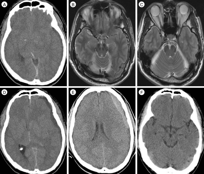
Brain computed tomography (CT) scans revealed an isodense left-sided CSDH with marked cerebral shifting (Fig. 1A). There was no evidence of source of this hemorrhage with temporal predilection on the CT angiogram. On magnetic resonance (MR) image subsequently obtained, the abnormal intensity within the subarachnoid space and the brain parenchyma was not visible. The patency without steno-occlusion in both transverse and sigmoid sinuses was clearly delineated on T2-weighted sequences (Fig. 1B, C). This patient has received a trephination and SDH drainage, after that he was sent home with resolution of headache. Approximately 2 weeks later, however, he developed an excruciating pain in the temporal and parietal regions with recurrence of subdural collection. The site and density of hematoma was similar to the first presentation (Fig. 1D). He was immediately returned for subdural irrigation and decompression through the prior burr-holes. The patient's clinical course was not eventful, but he complained of a mild headache again. Follow-up CT scanned just prior to discharge was strikingly for the newly-formed thin hematoma at the operative site (Fig. 1E). Another evacuation of this subacute subdural clot was not deemed to be necessary.
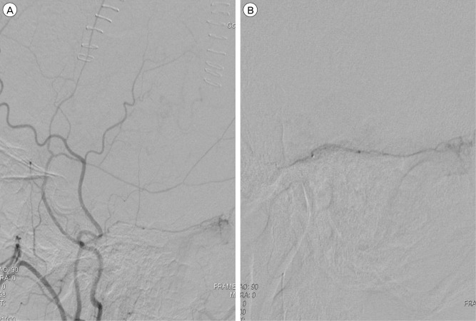
At this time, an active intervention was sought for this patient who had an intractably recurring CSDH. On the 7th day after the second surgery, angiography was performed to rule out an occult vascular lesion. A flow-guided type microcatheter (Prowler 10Ōäó, Cordis Neurovascular, Miami Lakes, FL, USA) was positioned in the main trunk of the MMA for selective angiography. The frontal and parietal branch of the MMA was appeared normally, and the abnormal membrane staining on the affected side was not detected. A left external carotid angiogram disclosed a dural AVF between the petrosquamosal branch of the MMA and the transverse-sigmoid sinus without retrograde cortical venous draining. The AV shunt had no connection to the internal carotid artery and its branches. It was suggested that the bleeding from the draining venous system of the dural AVF led to refractory CSDH. The microcatheter was introduced into the petrosquamosal branch of the MMA, thereafter polyvinyl alcohol particles ranging 150 to 250 ┬Ąm were distally injected (Fig. 2A, B). After trans-arterial obliteration of the feeder and fistula, the AV shunt disappeared. The recurrent hematoma of this patient did not increase, and his complaints of headache gradually subsided. The brain CT at one year following the embolization therapy revealed complete regression of the subdural hematomas (Fig. 1F).
DISCUSSION
Intracranial dural AVFs are pathologic communications between dural arteries and dural venous sinuses, meningeal veins, or cortical veins. These uncommon conditions are usually located in the transverse, sigmoid, cavernous, and superior sagittal sinus.2) The characteristic imaging feature is thrombosed sinus, dilated vessel, and hemorrhage within the adjacent cerebrum.17) In the present case, angiograms indicated the existence of dural veins that were connected to the anomalous artery arising from the left MMA at the lower posterior convexity. Therefore, the author made a firm diagnosis of a dural AVF in the area of the transverse-sigmoid sinus, and it was assumed that the oozing from the route of drainage with high flow inputs was responsible for the progression and relapse of CSDH.16) Another explanation for refractory CSDH is that repeated bleeding into the subdural space caused by the rupture of neo-vessels in an external membrane of hematoma.7)10)13) However, angiography of this patient did not visualize cotton wool-like staining along the branches of the MMA, which is microcapillary network of the hematoma membrane.
Dural AVFs have various clinical manifestations from aggressive neurological defects to no or minor symptoms and signs depending mainly on the pattern of abnormal drainage of the veins.1) Classification systems which are based on its mode of venous flow are used in cases of DAVF to estimate hemorrhage risks and to decide management strategies.4) Intracranial hemorrhage occurs in less than 20% of all these pathologies, and the bleeding is usually subarachnoid, more infrequently intra-parenchymal, and rarely in the subdural space.19) For that reason, the presence of dural AVF was not initially considered in the author's case with only CSDH without ICH and SAH. In the pertinent literature, there have been only 4 documented cases with idiopathic dural AVF complicated by acute non-traumatic SDH.5)6)7)8) Dural AVFs near the superior sagittal sinus were confirmed by cerebral angiogram in all of them. The sinus was well delineated as the drainer in those cases and in this report; however, angiography did not show a retrograde drainage into the cortical veins in the vicinity of the fistulas. The initial complaint was acute headache in the five patients including this representative case, but this is a unique illustration of the case with dural AVF that complicated pure CSDH.3)
Non-traumatic CSDH is an uncommon pathology in neurosurgery, and its diagnosis and treatment is a not straightforward process. Neurosurgeons must be aware of the differential diagnosis for spontaneous CSDHs because those develop secondarily to a few kinds of vascular lesion, such as aneurysms, arterial rents, arteriovenous malformations, intracranial AVFs, and veno-sinus thrombosis.8)9)15)18)21) According to literature review, a few cases with SDH or effusion, an exclusive revelation of cerebral veno-sinus thrombosis, have been reported thus far.10) It has been proposed that the formation of SDH in this rare occurrence was associated with increased venous and capillary pressure.11) Both CT and MR scans with arterio-venography are necessary to detect an occult vascular lesion and to prevent future problems in patients with a non-traumatic CSDHs. In addition, conventional angiography is the most valuable and decisive means for such patients and both internal and external carotid injections are essential for diagnosis and therapy. This briefing implies that dural AVF might be the cause of rare cases with intractable CSDH that were cured by the craniotomy and extensive membranectomy. Consequently, angiography should be recommended for intractable CSDH before such an aggressive surgery.
Craniostomy and intravascular approach provides the least invasive and definitive treatment for the rare condition of CSDH and coexisting dural AVF. Trans-venous embolization, which is one of the most effective therapies for dural AVFs, is now considered as an alternative to the craniotomy and excision in many cases.14) Trans-arterial embolization may be beneficial to another patients had a dural AVF, although it often lead to incomplete occlusion of the venous pouch.20) In the present case, however, the AVF can be secured by trans-arterial approach through the left MMA, because it was not difficult to select and penetrate the dural feeder in a single session. During the trans-arterial procedure, liquid embolic materials should penetrate into the draining veins to obliterate the fistula permanently.12) For this illustrative case, after the super-selective advancing a microcatheter to the draining vein, the interventionist administered polyvinyl alcohol particle into the fistula and veins, and the dural AVF was completely eliminated.
CONCLUSION
The author reported on a non-traumatic case who initially presented with CSDH caused by bleeding from a dural AVF involving the transverse-sigmoid sinus. Brain investigation including dynamic MR image and CT venography and catheter angiography is strongly recommended for the cases with acute SDH and intractable CSDH of obscure origin.
 PDF Links
 PubReader
 Full text via DOI
 Full text via PMC
 Download Citation
 Print
Print


