Migrated coil and damaged stent removal during coil embolization, using an additional, retrievable stent: A case report
Article information

Abstract
One of the common complications that can occur during coil embolization of cerebral aneurysms, is migration of coil lump alone. The removal of these migrated coils has been reported on a few occasions. On the other hand, rare complications would include the migration of the coil with subsequent stent dislocation. Currently, there is no standardized method to correct the complications of stent dislocation, and very few instances of this complication have been reported previously. In this report, we introduce a case of coil migration combined with stent dislocation. This occurred during coil embolization of an unruptured aneurysm of the distal, left internal carotid artery for a 52-year old woman. We retrieved both the damaged stent and migrated coil using another retrievable stent successfully with no more further complications. In the present report, we describe in detail how we corrected the complication successfully stent, and we discuss why this rescue maneuver is reasonable option for the complication mentioned above.
INTRODUCTION
One of the common procedure-related complications of endovascular coil embolization is coil migration [3]. If not retrieved, the migrated coil could induce further complications, including thromboembolic events [1,7]. Despite the lack of standardized guidelines for the treatment of coil migration, there is an abundance of such clinical cases that have been reported [4,6,8,11].
However, coil migration complicated with stent dislocation is extremely rare, and very few such cases have been reported [2]. We experienced this rare and challenging complication during endovascular coil embolization. In the following report, we describe the method that we used to recover the migrated coil and dislocated stent. We also review some previously reported clinical cases with similar complications and discuss their clinical outcomes.
CASE DESCRIPTION
A 52-year-old woman was admitted for coil embolization of an unruptured aneurysm. Transfemoral cerebral angiography (TFCA) prior to the procedure revealed an aneurysm in the left distal internal carotid artery (ICA). The aneurysm was relatively small but seemingly increasing in size; thus, we decided to treat it. A 6F Sofia intermediate catheter (MicroVention, Tustin, California, USA) was placed on the left cavernous ICA under guidance of 7F Arrow sheath (Arrow International LLC, Chihuahua, Mexico). Initially, with the guidance of a micro guide wire (Synchro, Boston Scientific, Natick, Massachusetts, USA), one microcatheter was placed on M1, and another one (pre-shaped 90, Excelsior SL-10, Boston Scientific, Natick, Massachusetts, USA) on the aneurysmal sac. We inserted a framing coil (Axium Prime 3D, 2.5 mm–4 cm coil, Medtronic, Irvine, California, USA) into the aneurysm, and noted a coil loop protrusion. As a result, we unfolded the stent (Acclino, 4 mm–25 mm stent, Acandis, Pforzheim, Germany) to cover the aneurysmal neck and inserted a framing coil using the jailing technique (Fig. 1).
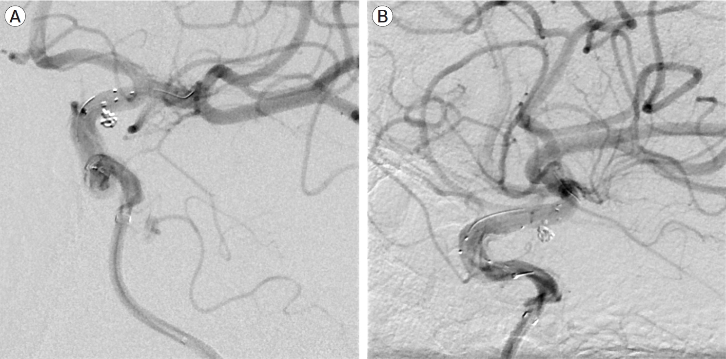
Frontal (A) and lateral (B) angiogram showing that initial coil embolization was successfully completed by stent application and jailing technique.
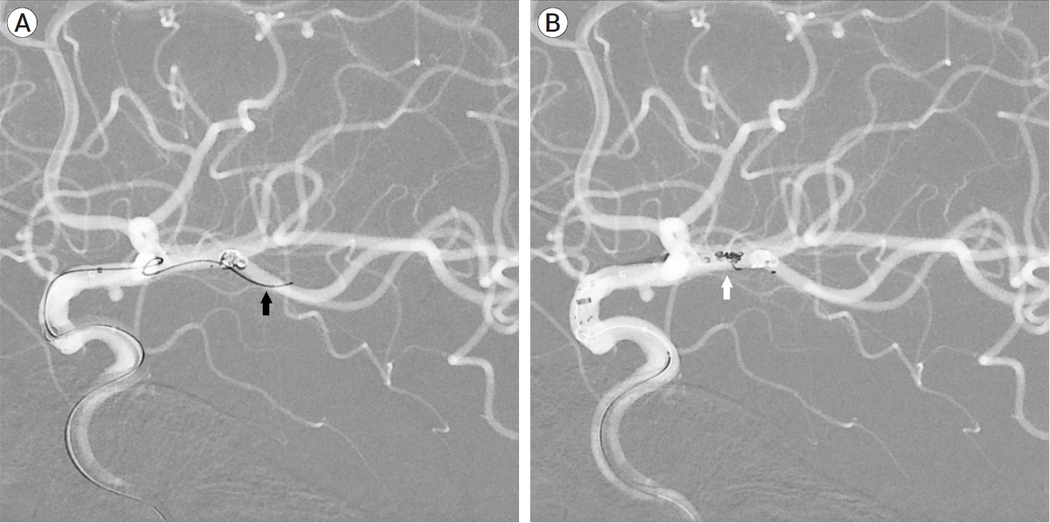
We began to retrieve the stent wire, but the stent shifted to the proximal portion, and the coil lump migrated to the distal portion of the middle cerebral artery (MCA) (Fig. 2). The stent constantly repositioned; thus, we removed both the migrated coil and the stent. We placed a microcatheter (Rebar 18, eV3, Irvine, California, USA) on the distal portion of the migrated coil. Then, we stabilized the coil and retrieved it using another stent (Solitaire Platinum, Medtronic, Irvine, California, USA) (Fig. 3). After shifting our attention back to the aneurysmal sac to perform the coil embolization, we inserted another framing coil (Axium Prime 3D, 2 mm–3 cm coil, Medtronic, Irvine, California, USA). We next attempted to retrieve the residual damaged stent. After several trials, we captured the damaged stent (Acclino stent, Acandis, Pforzheim, Germany) using the additional stent (Solitaire Platinum) and removed both successfully (Fig. 4).

Frontal (A) and lateral (B) road map view showing the migrated coil to M1 (black arrow) and the proximal end (white arrow) and distal end (red arrow) of dislocated stent in the proximal portion of the ICA. ICA, internal carotid artery
During the retrieval, another microcatheter (black arrow) was placed on the distal portion of the migrated coil (A), and the migrated coil lump (white arrow) was retrieved (B) using another stent.
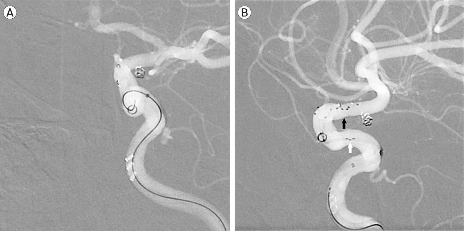
After coil embolization, another attempt to retrieve the damaged stent was made (A). The damaged stent (black arrow) was captured using another stent (white arrow) (B) and retrieved successfully.
On the angiogram that followed the procedure, blood flow at the distal portion of the aneurysm remained intact, with no dissection or extravasation (Fig. 5). The patient remained stable during the procedures and developed no subsequent neurological complications.
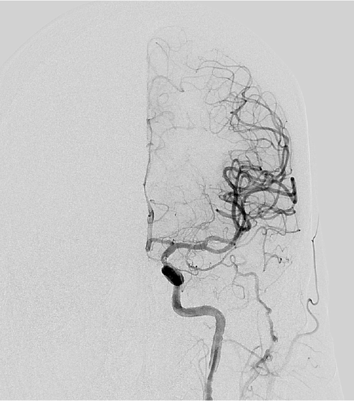
Final angiogram of left ICA showing intact blood flow and no further complications, such as dissection or extravasation, were noted. ICA, internal carotid artery
DISCUSSION
Coil migration and stent dislocation during coil embolization for the treatment of intracerebral aneurysm at the same time is a rare but possible complication. There are some known risk factors that increase the possibility of stent migration, such as tortuosity of vessel, irregularity of vessel caliber, thrombotic or calcified change of vessel, inadequate diameter of stent and excessive forward force during stent deployment [2,9]. When deploying a stent, an operator should consider risk factors mentioned above and make an effort to prevent the complication.
But when this desperate complication occurs, a migration of an intravascular stuff, a rescue maneuver should be considered to prevent serious complication expected to occur subsequently. Thromboembolic events due to migrated coil meshes are amongst common complications; thus, it is reasonable to remove these when migrated, to prevent such events [1]. While withdrawing the microcatheter used for deploying the stent, we noticed some resistance. Initially, we thought that this was due to partial stent deployment and an anchor being stuck in the catheter (Fig. 6). It was reasonable to assume that the stent migrated proximally, when pulling the stent catheter. Then stent was damaged due to constant reposition after dislocation. Successful removal of the migrated stent has been previously reported [9] and we therefore attempted to retrieve the migrated stent. Successful recovery would prevent further thromboembolic events without the need for antiplatelet agent administration.
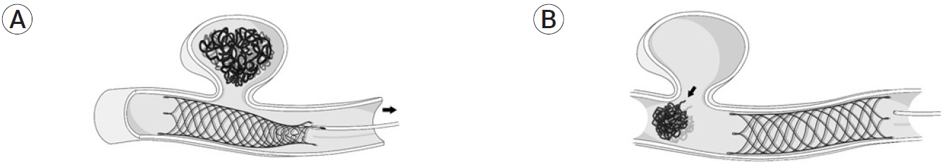
Schematic approach supporting comprehension of the reason why a deployed stent was damaged and migrated. One anchor of the stent was potentially stuck in the catheter, the stent migrated when pulling the catheter (arrow) (A), and coil migration followed as neck of the aneurysm was uncovered (arrow) (B).
Procedure-induced complications should always be taken into account when retrieving migrated intravascular devices. We performed a literature review of similar cases to learn about previously used treatments for the specific complication. Authors reporting the same complication were identified (Table 1) [5,9,12]. In this case, Vora et al. [12] reported a stentectomy of a migrated stent (Neuroform, 4.5 mm–30 mm stent, Natick, Massachusetts, USA) using an L5 Merci Retriever. However, extravasation occurred during the procedure and the patient was declared brain dead.

Summary of prior reported rescue methods for cases with the complication of stent migration
An animal study on a porcine model attempted 24 trials of Solitaire stent removal using 3 different stent retrievers (Trevo ProVue; EmboTrap II revascularization device; 3D revascularization device) [10]. The study reports successful stentectomy in all trials and no further procedure-related complications. Furthermore, the study suggests that an open tubular design of the Solitaire stent would allow for successful rescue maneuver and prevent further procedure-induced vascular injury.
Considering the aforementioned study, careful selection of a device and technique for the retrieval procedure a safe stentectomy can be made possible. When retrieving a migrated stent, the operator should take the type of the stent into account, to select the most suitable retrieving device. Commonly used devices include the snare device, Merci retriever, and retrievable stent [2,5,9,12]. Snare and Merci retriever are commonly used for damaged stent retrieval during mechanical thrombectomy in ischemic stroke. In the present case, we used a retrievable stent to recover a migrated, damaged stent during endovascular coil embolization of an unruptured intracranial aneurysm. It is important to consider the possibility of stent interaction when using a retrievable stent to retrieve another stent. For example, Vora et al. reported a case in which an open-cell type stent (Neuroform stent) was removed by a Merci retriever, but extravasation of contrast was noted [12]. In contrast, we removed a closed-cell type stent (Acclino stent, Acandis, Pforzheim, Germany) using another closedcell type stent (Solitaire Platinum). A closed-cell type stent is likely to be more easily retrieved by a single stent stentectomy because the struts of such stents move in an integrated way.
Operators should also select a proper rescue technique. There are some well-known techniques such as ‘deploy and engage’ and ‘loop and snare’ [9]. We deployed a retrievable stent (Solitaire Platinum) through the strut of a damaged stent. Both were retrieved simultaneously, similarly to a mechanical thrombectomy for ischemic stroke using a single stent. Our technique was technically similar to ‘deploy and engage’, but differed slightly as we passed the retrievable stent through the strut of damaged one. Contrary to the classic ‘deploy and engage’ technique, ours made it possible to retrieve even an open-cell type stent.
CONCLUSIONS
The migration of a coil and dislocation of a stent is rare but possible. There are few reported clinical cases of complication, and the technique for treatment is non-standardized. More clinical trials are needed to establish standardized management for this complication. However, the selection of a proper device and technique for rescue maneuver, it is more likely to overcome the complication without further issues. It is therefore important for operators to be familiar with various devices and techniques. Based on the present case, using a retrievable stent to remove the damaged stent, both stents of the closed-cell type, is a considerable option for this rare complication.
Notes
Disclosure
The authors report no conflict of interest concerning the materials or methods used in this study or the findings specified in this paper. The patient provided written informed consent for this publication.