


INTRODUCTION
Tetralogy of Fallot (TOF) is a congenital abnormality which occurs during development of cardiac vasculature, which occurs in 2.0-3.6 per 10,000 live births. Its combination with anatomic variations of the aortic arch and adjacent branches have occasionally been reported [2,8].
Proatlantal intersegmental artery (PIA) itself is also a rare congenital abnormality of cerebrovascular development [3]. PIA requires no special treatment, but may be associated with cerebrovascular disease, such as aneurysm, due to abnormal cerebral hemodynamic changes unto primitive vasculature.
Here, we presented a case of intracranial aneurysm combined with the aforementioned congenital variants, which has not been reported to our knowledge. For this patient, endovascular treatment can have elevated risks, however they are likely lower than open surgery. Thus, flow diverter placement may have an advantage in treating wide-necked aneurysms in patients with complex cardiocerebral disease.
CASE DESCRIPTION
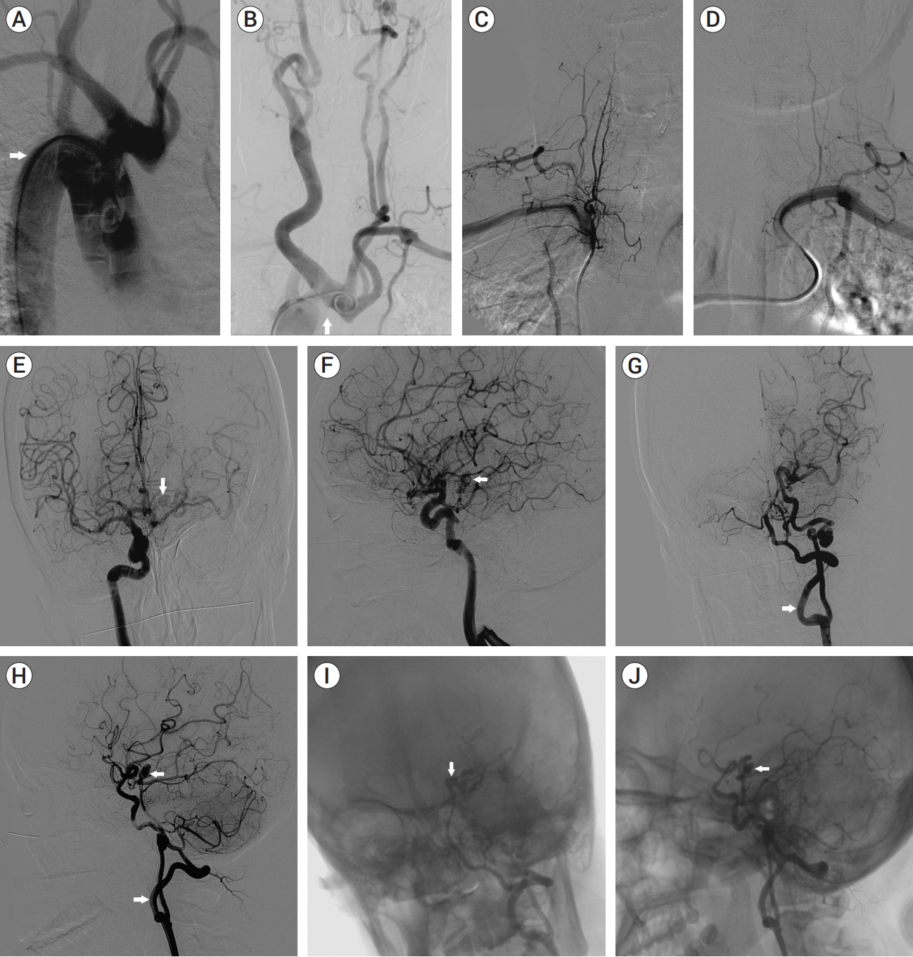
A 41-year-old female presented with persistent light-headedness and fatigue for 20 days. Family history was non-contributory. She reported longstanding mild limitation of physical activity. Physical examination showed cyanosis of the lips and longstanding diastolic murmur. Laboratory tests showed that the arterial blood oxygen partial pressure was 52 mmHg, oxygen saturation 87% and hemoglobin was 184 g/L. Echocardiography confirmed TOF. Cranial magnetic resonance imaging (MRI) demonstrated a left posterior cerebral artery (PCA) aneurysm and showed remote cerebral infarctions in the right frontal and parietal lobes (Fig. 1). Digital subtraction angiography (DSA) was further performed, confirming P1 segment aneurysm and right aortic arch (RAA). Also, the right common carotid artery (CCA), left internal carotid artery (ICA), left external carotid artery (ECA) and left subclavian artery (SA) all branched from a common main trunk from the aortic arch. Bilateral vertebral artery (VA) were absent, and type I PIA of left side was observed (Fig. 2).
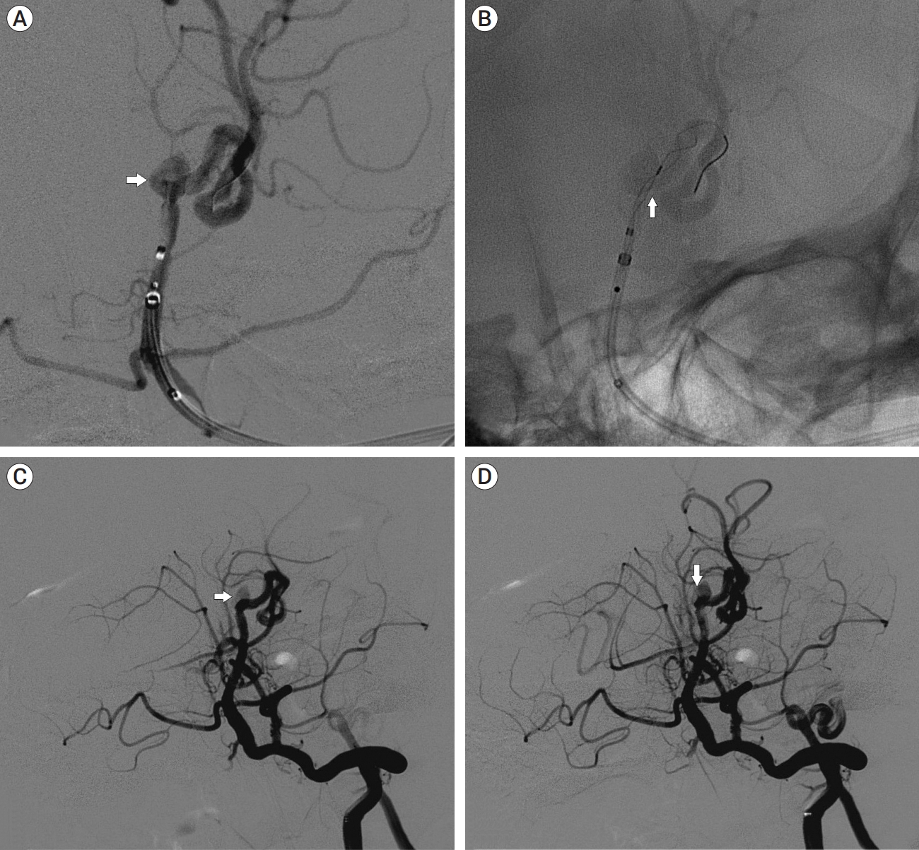
Nasal prong oxygen was given. After multidisciplinary discussion, aneurysm treatment with a Tubridge flow diverter (FD) (4.0×45 mm) was decided. Oral dual-antiplatelet medications (aspirin 100 mg/d aspirinand clopidogrel 75 mg/d) were prescribed 7 days before the procedure. The procedure was performed under general anaesthesia and via a transfemoral approach. Heparin (4000U) was administered. The tip of a long vascular sheath was placed in left ICA. An intracranial support catheter was then transferred to the basilar artery via left PIA, and a microcatheter guided by a microwire was then delivered to P2 segment of left PCA. The FD was implanted into the parent artery (P1 segment of PCA) through the microcatheter (Neuroslider 27 catheter for 4.5 mm and 5.5 mm devices and Neuroslider 17 catheter for 3.0 and 2.5 mm devices).
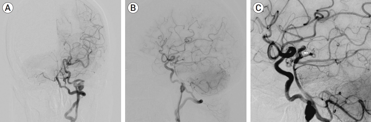
Immediate intraoperative angiography confirmed decreased contrast filling into the aneurysm sac after FD placement (Fig. 3). The patient continued high-flow oxygen and antiplatelet therapy after the intervention. Her dizziness resolved and there was no new discomfort. She continued home oxygen and oral dual-antiplatelet medications after discharge. Follow-up DSA at 6-months showed the aneurysm was not visible and likely obliterated (Fig. 4). Oral antiplatelet medications were adjusted to a single agent for long-term stent patency.
DISCUSSION
TOF is hypothesized to be caused by dysplasia of the infundibulum or conus of the right ventricle and can be accompanied by other large vessel anomalies such as the aortic arch itself [4,7]. There are rare reports of combinations of TOF and RAA or other anatomic variants. Cerebrovascular anomalies and congenital cardiac abnormalities may be derived from embryonic development [5,6]. Most patients with TOF are diagnosed and treated during infancy, and only 3% of patients over 40 years old remain untreated. In this case, cardiac surgery was not performed in childhood as symptoms were mild. However, TOF itself may be an important cause of cerebrovascular events, which may be due to secondary to erythrocytosis, activation of procoagulant pathway induced by hypoxia/hypoxia, etc. Thus, an ischemic event is more commonly seen [1].
PIA is the residual manifestation of original fetal carotid-vertebral-basilar anastomosis during embryonic development. Sato et al. and Nonaka et al. have both reported cases combinations of PIA with intracranial aneurysm [6]. It has been reported that 59% of PIA patients are complicated with other cerebrovascular abnormalities, and approximately 10% of these are intracranial aneurysms. Arteriovenous malformation and vein of Galen malformation are also related to PIA. All of these could suggest hemodynamic changes caused by anatomic variants, such as PIA, may play an important role in the occurrence of cerebrovascular diseases. An association between TOF and PIA has not been definitively established, however Ojha et al. suggest the association is likely under-reported [7].
In this case, extensive bleeding and/or fluid rehydration during open surgical clipping may increase the risk of heart failure in the context of intraoperative blood volume changes, which is often catastrophic in patients with TOF. In addition, vasoactive agents, such as norepinephrine, may induce pulmonary hypertension and subsequently worsen right ventricular function [8]. Therefore, endovascular treatment would seem to be more appropriate. However, vessel tortuosity and stenosis in these cases are nevertheless obstacles and undoubtedly increase the difficulty of endovascular intervention. Traditional stent-assisted coiling was not chosen as dual microcatheters technique would be required, mandating bilateral access. By contrast, FD employs a single microcatheter and entails a shorter procedure. The wide neck was felt to preclude simple coiling, particularly in a congenitally arteriopathic procedure.
CONCLUSIONS
Congenital cardiovascular diseases may also be complicated by cerebrovascular abnormalities. Non-invasive cerebrovascular screening should be considered. Cerebrovascular investigation revealing aneurysm or other cerebrovascular diseases in the context of congenital cardiac disease may require DSA for detailed assessment. For wide-necked intracranial aneurysms with complex cardio-cerebrovascular variations, FD may be a comparatively safe treatment choice in the modern endovascular era.

 PDF Links
 PubReader
 ePub Link
 Full text via DOI
 Download Citation
 Print
Print


